Cardiac MRI vs. CT Angiography: When Each Matters
Understand the differences between cardiac MRI and CT angiography, when each is most useful in preventive cardiology, and how these tests work together to detect early heart disease and guide personalized care.
.svg)
.svg)
In preventive cardiology, no single test tells the whole story of your heart health. Each imaging tool provides a different window into how your heart and blood vessels are functioning and how they may be changing over time.
Rather than asking which test is “better,” the more meaningful question is which test is most appropriate for you at a given moment. A longitudinal approach tracking trends rather than relying on a single snapshot allows Cardiac MRI and CT Angiography to be used strategically and complementary in truly personalized preventive care.
The Power of Trends (MRI vs. CT Over Time)
A single cardiac image can reveal important information, but repeated assessments over time often tell a much deeper story.
For example, two individuals may both have early plaque visible on CT Angiography. One may show stable plaque over years, while another may demonstrate progression suggesting very different risk trajectories. Similarly, two people may have normal heart function on Cardiac MRI, but subtle changes in myocardial tissue or function over time may signal early disease in one and stability in the other.
CT Angiography is particularly powerful for tracking structural changes in coronary arteries, such as plaque burden and progression. Cardiac MRI excels at tracking functional and tissue-level changes, such as heart muscle performance, scarring, inflammation, and blood flow.
Together, trends across both modalities provide a richer, more accurate picture than either test alone.
Establishing Your Cardiac Baseline
To use these tools effectively, we first establish a clear cardiac baseline during periods of good health. This allows future imaging to be interpreted in context, making subtle changes easier to detect and understand.
Key baseline cardiac assessments may include:
- Coronary artery calcium (CAC) scoring or CT Angiography
- Cardiac MRI for heart structure and function
- Comprehensive lipid and inflammatory blood panels
- Blood pressure and metabolic risk assessment
- Functional testing where appropriate
The ideal time to establish baselines is during your 30s or 40s, before age-related changes begin to accumulate. However, it's never too late to begin, even later baselines provide valuable reference points for detecting future changes.
Personalized Monitoring Intervals (Choosing MRI vs. CT Over Time)
Not everyone needs the same imaging at the same frequency.
If your initial CT Angiography shows minimal plaque and low risk, follow-up may focus more on labs, lifestyle, and periodic reassessment rather than repeated CT scans. If early plaque is detected, targeted monitoring may be recommended.
Similarly, if your Cardiac MRI shows excellent heart function, you may only need periodic reassessment. If subtle abnormalities are present such as early fibrosis or reduced blood flow closer follow-up may be appropriate.
We tailor imaging schedules based on your initial findings, family history, and evolving risk factors. Your plan adapts as your health evolves.
Technology-Enhanced Tracking: What Each Modality Does Best
Modern cardiac imaging allows for highly precise, quantitative tracking of heart health.
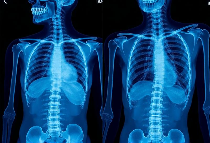
CT Angiography — Best for Coronary Anatomy
CT Angiography provides high-resolution images of your coronary arteries, allowing physicians to:
- Visualize plaque location and burden
- Assess degree of arterial narrowing
- Track plaque progression over time
- Identify high-risk plaque characteristics
It is particularly useful for detecting early atherosclerosis before symptoms develop.
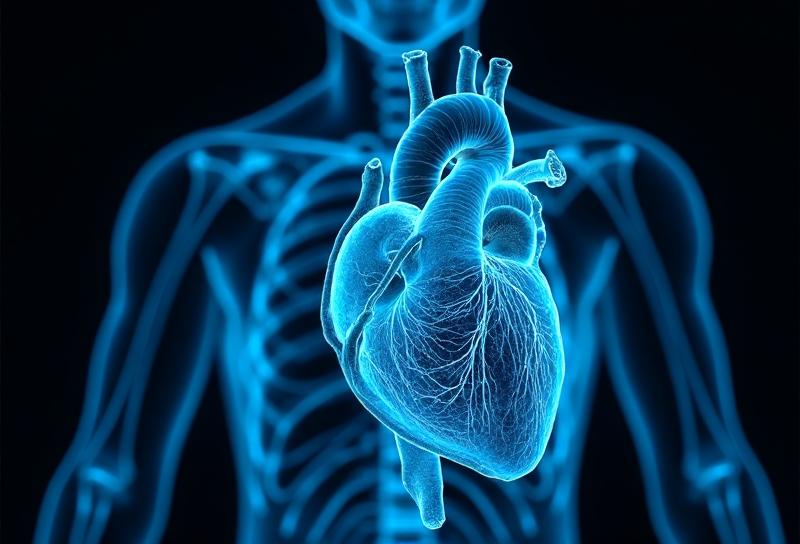
Cardiac MRI — Best for Heart Function and Tissue Health
Cardiac MRI offers unmatched detail in assessing how your heart actually works, including:
- Heart muscle strength and efficiency
- Blood flow to the heart (perfusion imaging)
- Detection of scar tissue or inflammation
- Evaluation of cardiomyopathies and structural heart disease
Where CT shows what your arteries look like, MRI shows how your heart is performing and why.
Beyond Imaging: Integrated Cardiac Data
A truly preventive approach does not rely on imaging alone. We integrate CT Angiography and Cardiac MRI findings with:
- Lipid panels and inflammatory markers
- Blood pressure trends
- Wearable data (heart rate variability, activity, sleep)
- Lifestyle and stress assessments
This integrated view allows us to connect structural findings on CT with functional insights from MRI and real-world health data — creating a complete picture of your cardiovascular trajectory.
The Value of Institutional Memory in Cardiac Care
We maintain detailed records of all your cardiac imaging and lab results, allowing direct comparison over time.
When you return for follow-up studies, our physicians can immediately compare your current CT Angiography or Cardiac MRI with prior results. This continuity makes it easier to identify meaningful changes — whether improvement, stability, or progression — rather than interpreting each scan in isolation.
This institutional memory is critical for truly effective preventive cardiology.
Actionable Insights: When MRI or CT Changes Your Care
The ultimate goal of using Cardiac MRI and CT Angiography in prevention is early, meaningful intervention.
For example:
- If CT Angiography shows increasing plaque burden, we may recommend more aggressive lipid management, dietary changes, or medication.
- If Cardiac MRI reveals early myocardial fibrosis or reduced perfusion, we may focus on optimizing blood pressure, inflammation, and metabolic health.
- If both modalities are stable over time, we may prioritize lifestyle optimization rather than medical intervention.
In this way, trends — not single test results — guide smarter, earlier, and more personalized cardiovascular care.
Related Articles
Understanding Coronary Calcium Scoring
Coronary calcium (CAC) scoring detects early signs of heart disease before symptoms appear, helping physicians assess risk, interpret your Agatston score, and create a personalized prevention plan.
.svg)
Why Full-Body MRI Isn't Always the Answer
A full-body MRI can sound reassuring, but it can also create false alarms. Learn why a more targeted, risk-based approach is often safer, clearer, and more effective for finding disease early.

The Longitudinal Approach to Health Monitoring
Tracking your health over time enables earlier detection of disease by revealing meaningful trends in imaging, labs, and risk factors so physicians can intervene sooner with personalized preventive care.
Your Defense Against Disease Starts Here
Schedule a complimentary PreScan Consultation to discuss how our clinically restrained, longitudinal approach differs from mass-market screening.
.svg)